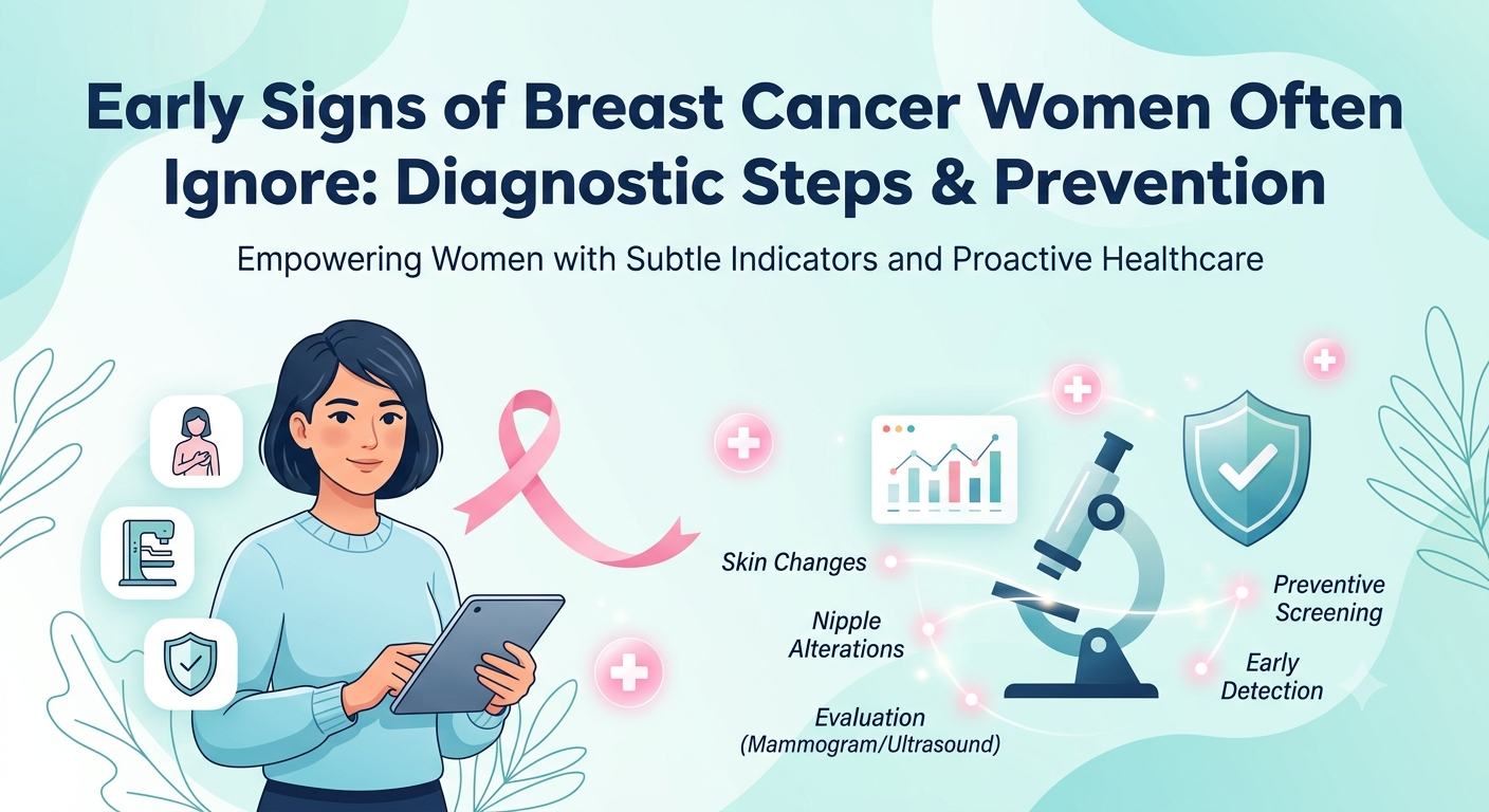
Early Signs of Breast Cancer Women Often Miss & Next Diagnostic Steps
By Dr. (Maj) A.P. Dubey in Medical Oncology Cancer
Sep 26, 2026
Beyond the Lump: Subtle Early Signs of Breast Cancer Women Often Miss & Next Diagnostic Steps
How do you spot the early warning signs of breast cancer before a lump appears?
Early warning signs often extend beyond a painless lump. Key symptoms include:
- Skin Texture Changes: Dimpling or pitting resembling an orange peel (peau d’orange).
- Nipple Alterations: Sudden inversion, flattening, or spontaneous clear/bloody discharge.
- Tissue Changes: Unexplained swelling, persistent localized pain, or enlarged underarm lymph nodes.
Early evaluation via Digital Mammography and High-Resolution Ultrasound is essential for accurate diagnosis.
Breast cancer is one of the most frequently diagnosed cancers in women globally, making early detection vital for effective treatment. However, with advances in modern oncology and diagnostic technology, early detection offers exceptionally high cure rates and successful treatment outcomes.
While many women actively look out for obvious symptoms like a hard, painless lump, several subtle physical and tissue changes often go unnoticed or are mistakenly attributed to hormonal shifts, aging, or minor skin conditions.
Recognizing these overlooked early warning signs, understanding the essential diagnostic pathways, and adopting proactive preventive strategies are critical steps in safeguarding your breast health.
1. Subtle & Commonly Ignored Early Signs
Beyond a noticeable lump, breast cancer can manifest through various subtle symptoms that require prompt medical evaluation:
- Changes in Skin Texture (Dimpling or Pitting): Skin on or around the breast that begins to look like the dimpled peel of an orange (peau d’orange) can indicate underlying inflammation or lymphatic blockage.
- Nipple Changes or Inversion: A suddenly inverted (pulled inward) nipple, asymmetry, or persistent flattening that was not previously present.
- Unusual Nipple Discharge: Spontaneous discharge—especially if it is clear, bloody, or occurring from a single duct—warrants immediate investigation.
- Persistent Localized Pain or Discomfort: While cyclic breast pain tied to menstrual cycles is common, non-cyclic, focal pain in one specific spot that persists throughout the month should be evaluated.
- Skin Flaking, Redness, or Scaling: Persistent itchiness, scaling, or redness around the nipple or areola that mimics eczema or dry skin and does not respond to topical creams.
- Swelling or Fullness in the Axilla (Underarm): Breast tissue extends toward the armpit. Enlarged or firm lymph nodes under the arm or near the collarbone can sometimes appear before a primary breast tumor is felt.
Note: Finding any of these symptoms does not automatically mean you have cancer, but it does mean your body is asking for a professional clinical checkup.
2. Essential Diagnostic Steps: From Screening to Confirmation
When an irregularity is noticed during self-examination or a routine clinical visit, a structured multi-tier diagnostic approach ensures an accurate and timely evaluation.
| Step | Phase | Key Diagnostic Actions |
| Step 1 | Clinical Evaluation | Detailed Medical History and Professional Physical Examination |
| Step 2 | Diagnostic Imaging | • Digital Mammography/3D Tomosynthesis: Primary screening • High-Resolution Ultrasound: Cysts vs solid masses • Breast MRI: High-risk screening & tissue mapping |
| Step 3 | Biopsy & Staging | • Core Needle Biopsy (CNB): Image-guided tissue sampling • Histopathology: ER, PR, and HER2/neu receptor profiling |
Key Diagnostic Tests Explained:
- Digital Mammography & 3D Tomosynthesis: The gold standard for detecting microcalcifications and non-palpable lesions long before they can be felt.
- High-Resolution Breast Ultrasound: Excellent for dense breast tissue and evaluating whether a lump is fluid-filled (a benign cyst) or a solid mass.
- Core Needle Biopsy: If an abnormal area is identified, an image-guided core biopsy extracts a tiny tissue sample to provide a definitive histopathological diagnosis, identifying receptors (ER, PR, HER2) to tailor personalized treatment plans.
3. Preventive Strategies & Risk Reduction
While certain risk factors such as age, gender, and inherited genetic mutations (BRCA1/BRCA2) cannot be changed, adopting evidence-based lifestyle habits significantly reduces overall risk:
| Prevention Pillar | Actionable Guidance |
| Monthly Breast Self-Examination (BSE) | Perform a 5-minute self-exam monthly (ideally 3–5 days after your period ends) to get familiar with your normal tissue texture. |
| Annual Clinical Screening | Women aged 40 and above (or earlier if there is a family history of breast or ovarian cancer) should schedule an annual screening mammogram and specialist consultation. |
| Maintain Healthy Body Mass Index (BMI) | Post-menopausal estrogen is primarily produced in fatty tissue. Managing weight through balanced nutrition reduces excess circulating estrogen. |
| Active Lifestyle & Physical Exercise | Aim for at least 150 minutes of moderate aerobic activity or 75 minutes of vigorous exercise weekly. |
| Limit Alcohol & Avoid Tobacco | Even low-to-moderate alcohol consumption is linked to a higher risk of hormone-receptor-positive breast cancers. |
Frequently Asked Questions (FAQs)
Q1: What are the early warning signs of breast cancer other than a lump?
Answer: Beyond a painless lump, early warning signs of breast cancer include skin dimpling or pitting (resembling an orange peel), sudden nipple inversion, unexplained clear or bloody nipple discharge, localized persistent breast pain, redness or scaling around the nipple, and swelling or firm nodes in the underarm or collarbone area.
Q2: Which diagnostic test is best for detecting breast cancer at an early stage?
Answer: Digital Mammography with 3D Tomosynthesis is the gold standard for early detection, as it can identify microcalcifications and tiny tumors long before they can be felt. For dense breast tissue, doctors often combine digital mammography with a high-resolution breast ultrasound or dynamic breast MRI for maximum accuracy.
Q3: At what age should women start getting screened for breast cancer?
Answer: Women with an average risk should begin annual screening mammograms at age 40. Women with a family history of breast cancer or genetic markers like BRCA1 or BRCA2 should consult a specialist to begin personalized screening earlier, typically starting between ages 25 and 30.
Q4: Does finding a lump in the breast always mean it is cancer?
Answer: No. Over 80% of breast lumps are non-cancerous (benign). Common non-cancerous causes include fluid-filled cysts, fibroadenomas, hormonal fluctuations, or infection. However, any new or changing lump must be promptly evaluated by a specialist using diagnostic imaging or a biopsy.
Q5: How is a breast biopsy performed, and is it painful?
Answer: A Core Needle Biopsy (CNB) is a quick, minimally invasive procedure where a doctor uses local anesthesia to numb the area and an image-guided hollow needle to collect a tiny tissue sample. Because the area is completely numbed, patients experience minimal discomfort.